The Most Famous Gram after Parsons

Gram_stain_01I love my bit of country rock, my wee daily dose of Americana. And ol’ Gram Parsons would be in the mix there, depending on the mood. But today I’m going to write about another Gram. A surname Gram, if you get my drift. And a man far removed from the world of the Byrds, Flying Burrito Brothers and cowboy boots.

Hans Christian Joachim Gram was a Danish microbiologist who could be described as the father of bacterial strain identification. For it was he who gave us the simple but effective staining method that has split the eubacterial kingdom right down the middle. After staining, a bacterium is either Gram-positive or -negative*.

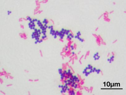
In 1884, Gram was working in the morgue of a Berlin hospital and wished to devise a stain that would colour up the bugs from sections of lung tissue and allow them to be discriminated from the background human cells. After some trial and error and a few modifications, we got what is now called the Gram stain. The stain works on the basis of dyes that react with the bacterial cell wall, which is composed of peptidoglycan. One group of bacteria have a thick layer of peptidoglycan, while another has a thin layer that is surrounded on the outside by a thick (and sometimes gooey) lipid membrane. The first stain applied during Gram staining, crystal violet, stains both types of cells purple. The next additive, iodine, is a mordant#, which in the case of the thick-walled cells fixes the crystal violet’s purple colour, but is occluded from the second group of cells by the lipid layer. Alcohol is then applied to the cells. This washes out the loosely held dye from the thin-walled cells, decolourising them, while having little effect on the thick-walled cells, which remain purple. A second colour (either safranin or fuschin, both red) is then added to the cells. This addition does not affect the thick-walled cells, which remain purple, while the colourless, thin-walled cells are now stained red. And so we have in a simple staining method two broad groups of cells distinguished on the basis of cell envelope morphology. Combined with other morphological features that can be seen under a reasonably high-powered microscope, the Gram stain yields a deal of information about the mystery bugs under investigation.

After isolating a bug, Gram staining is the first port of call for a bug hunter. A bug being Gram-positive or -negative allows us microbiologists to rule our mystery organism out of belonging to about 50% of the eubacterial kingdom. Whatsmore, in taking a gawk at our isolate under the microscope to check for Gram status we also get to cop a look at cell morphology. Is the critter a rod? Or a coccus (round)? Is it boat-shaped? Sausage-shaped? Are there spores? Do the cells occur in bunches of grapes? Or in chains? Do the cells exist as tetrads? Et cetera. Us bug hunters then follow a protocol to narrow down the possibilities of what our isolate might be. We test for the presence of enzymes such as catalase and oxidase. We test for the bug’s ability to metabolise a host of carbon sources. And finally, if we want to be really precise, we sequence the blighter’s DNA and compare it to a database of previously identified isolates.

Thus, the modern microbiologist is using a mixture of techniques, some of which date from the nineteenth century, and some which were invented last year. And even in this day and age of next-generation sequencing and CRISPR and aptamers all that, it’s still not time for Gram to sling his guitar!


*There’s a couple of “buts” and “howevers”, however. Some strains are Gram-variable. Depending on culture age or physiological state, they can be positive or negative. But these are outliers in a very successful and venerable classification system.

#A mordant is a substance that combines with a dye or stain and thereby fixes it in a material.

Some Good Sporeformers

Sporeforming bacteria have a bad rep. The food industry hates them; there are entire conferences dedicated to the elimination of Bacillus and Clostridium from milk powders and infant formula. Doctors and hospital managers quake at reports of high levels of C. difficile in their hospitals. There’s nothing to instill more fear in the general public than talk of an anthrax outbreak, be it related to farm animals or bioterrorism. It is spores’ extreme resistance to all the usual treatments that we use to eliminate bacteria, combined with their ability to survive for years in a dormant state that causes our fear and loathing of them. But we must not forget that, in the case of certain sporeformers, these properties can be harnessed by biotechnologists. Not all spores are villains.

The first sporeforming good ‘un that I came across in my study of microbiology all those years ago was Bacillus thuringiensis. Many readers will have encountered the terms “Bt” or “Bt proteins” or “Bt crystals”, and perhaps some of you will have applied a product containing these terms in its list of ingredients to a cabbage patch or a flower bed. B. thuringiensis spores contain protein crystals called δ-endotoxins which have insecticidal action and have been used for a hundred years as biocontrol agents for the caterpillars of moths and butterflies. You spray your field with a preparation of Bt spores. When the caterpillars get a-munching they ingest the spores whose protein crystals then proceed to turn the poor wigglers’ insides into mush. The great thing about applying Bt spores to your land is that they persist in the soil for years. They don’t wash into the groundwater like conventional chemical pesticides, and they do not cause any damage to the environment or the man or woman charged with applying them. Bt preparations are the insecticide of choice for organic farmers. Genetically modified crops containing the gene sequence for a Bt protein, and which therefore produced endogenous Bt protein, were a cause celebre in the GM wars in the 1990s. I’m not going there!

Another triplet of sporeformers are being looked at as natural pesticides which would see use similar to Bt. Lysinibacillus sphaericus (formerly Bacillus sphaericus — these crazy taxonomists can’t leave any poor bug in peace!) is a mosquitocidal bacterium which harbours its insecticidal crystal in a mesh-like parasporal envelope and could be applied to swamplands close to towns and villages to reduce the impact of malaria-carrying mosquitoes. Imagine the lives that could be saved by such a simple and safe intervention. Pasteuria penetrans is a parasite of root-knot nematodes — pests to which are attributed 5% of annual global crop loss. The spores have been commercialized as a biological control agent. A cousin of P. penetrans, P. ramosa infects water fleas of the Daphnia genus. Infected hosts are completely sterilized and have a reduced life span.

Now, it’s time to describe a more esoteric and technical use for bacterial spores. Spores of the genus, Bacillus, all display a protein on their outer surface, the exosporium, called BclA. Genetic engineering of this protein — splicing a gene from another species onto the anchor region of BclA — allows scientists to stick any protein they want on the outside of these spores. What use is this, you may ask. Because of spores’ unique stability in the environment and inside living organisms, as well as their amenability to mass production processes such as freeze drying and inclusion in “wet” formulations, bioengineered spores offer huge possibilities. Recombinant protein-decorated spores can be purified and used as microparticles for industrial, bioremediation, or vaccine applications. Imagine a Bacillus spore genetically modified to present an antigen from the malaria parasite on their surface. You have a candidate for a malaria vaccine there. What’s more, an orally delivered vaccine (spores will survive passage through the low pH environment of the stomach). Because it has been shown that enzymes retain their activity when tethered to the exosporium, GM Bacillus are ideal for a range of biocatalytic processes; the clean-up of oil slicks, waste water treatment or the manufacture of bioethanol.

Anthrax and Argi(nase)-bargy

I’ve p2pha_humanarginasereviously referred to the evolutionary toing and froing between microbes and their hosts as an “arms race“. A microbe develops a sneaky new way to attack or evade its host. A couple of millennia later the host species evolves a way to overcome its foe. A new strain of the dastardly bug crops up, replete with a mutation that allows it slink past its host’s beefed-up and shiny new defences. The host species finds a way to block this latest ruse. Et cetera, et cetera. And so the deadly dance escalates.

The more you learn about disease-causing microbes (or, to be fancy, pathogens) the more one comes to appreciate the extent to which evolution has honed these suckers into becoming laser-guided killers. It would seem as if every single gene in their genome, every single structure from wall to flagellum — heck, every single molecule they possess — is purposed with doing damage to their host in order to win the ageless battle between pathogens and us decent folk.

Let’s take one example — that of Bacillus anthracis and one (just one of many!) of the weapons in its disease-causing arsenal (technically known as virulence factors): arginase. Everybody knows that B. anthracis is the causative agent of the deadly disease anthrax. It forms resistant spores that can persist in the environment for centuries and which can be freeze-dried and stored as a powder, making it the bug of choice for back room bio-terrorists. In a nutshell, B. anthracis infects the body by worming its way into cells, hiding out there, dividing, spreading to uninfected cells, and of course producing its lethal toxins. It (or evolution) has developed a myriad of ways to hide from or counteract the immune system. Arginase is just one of these.

One of the main cells charged with the elimination of B. anthracis and other invaders from the body is the macrophage. In Greek, macrophage means “big eater”, which pretty much encapsulates what these immune cells do — they travel around the body gobbling up bugs. But they don’t use teeth to masticate the microbial malfeasants — they use enzymes and chemicals to digest and obliterate them. One of the most effective chemicals employed by macrophages to scuttle invading bugs is nitric oxide, which acts on them in the same manner as a solar wind on a spaceship without deflector shields. Fizzle, fizzle, fizzle! Not very many bugs can withstand a blast of the old NO, and not very many bugs have developed a defence against it. Except, that is, B. anthracis.

Which is where arginase comes in. B. anthracis spores have significant amounts of the enzyme in their outer layer (the exosporium). The arginase competes with the macrophage’s nitric oxide synthase (the enzyme responsible for making NO) for its L-arginine substrate, snatching the cell’s raw materials before it gets the chance to stock up on the gas. No NO means the spores and the vegetative cells that germinate from them are spared a blast of solar wind and so have a higher chance of survival and reproduction. And killing us.

What better example of the immunological arms race could there be? One bug, one host, one defence mechanism (NO) and one way around it (arginase).

What is a Type Strain?

At the root of almost any microbiological study is a type strain. Lay people always ask what this is. In brief, a type strain is a particular strain (or breed) of a microbe that presents all of the characteristics that define a species. A perfect specimen, if you like. Using a type strain allows the microbiologist to perform studies on a particular species that will have relevance to all of the (sometimes thousands) of strains that make up that species. In my work on Bacillus cereus, an age ago, I used a couple of type strains in order to show that the staining techniques I developed would apply to the majority of B. cereus isolates that might be found in rice or milk or lasagna. It would have been folly to isolate my own bugs from a slice of ham from the local deli and then try and extrapolate that particular bug’s heat resistance, for example, to B. cereus stains that find their way into food all around the world.

Let’s extend the type strain concept to human beings. A perfect specimen person should display all the characteristics that define the what being Homo sapiens means: (s)he should walk upright, have 32 teeth, neither be a dwarf nor a giant, not be a diabetic, have a blood pressure within the normal range et cetera, et cetera. The perfect specimen should also be clonable and be amenable to cryopreservation so that they can be bulked up and sent around the world to participate in studies! And be genderless — just like bacteria. In other words, human type strains do not exist, and thus the execution of clinical trials and the like involving real people are peppered with layers of complexity and issues of extrapolation to the general population that us microbiologists don’t have to deal with.

Just as is the case with the classification of bacteria, the designation of a particular strain as the type strain is controversial. You come across grand debates in the scientific journals arguing why one wee fellow should be defrocked of his status as type strain, and another cheeky lad handed the crown. The type strain in question may not display the salt tolerance typical of the entire species, or may have a biochemical pathway untypical of the rest of his brethren. He may munch pentose sugars and spit out acetone, whereas the rest of the strains making up the species might turn their noses up at five-carbon sugars, and find nail polish remover inimical. That’s why when I do my studies, I like to have a couple of type strains. Just in case one of the wee fellows is about to lose his title. I also like to have some local isolates — fellows I’ve hunted down myself, grown up in the lab and done the necessary tests on to prove that they show the defining traits of the species. Using isolates from the particular foodstuff or environment that I am studying give the study more robustness.

The rules that govern the designation of a strain as a type strain are laid out very solemnly in a document called the International Code of Nomenclature of Bacteria. Very often type strains were defined in the early twentieth century and have stood the test of time, which is a tribute to the precision and diligence of previous generations of microbiologists.

An obvious question is: how do microbiologists get their hands on type strains? Well, just as there are seed banks all around the world where seeds are stored for many reasons, only one of which is the frightening scenario of the post-apocalypse re-seeding of the planet, there are collections of type strains. The most well known of such bodies is the ATCC — the American Type Culture Collection — which started out in the McCormick Institute in Chicago but is now located in Manassas, Virginia, and boasts 18,000 sq ft and 200 megafreezers of storage. The main European culture collections are the DSMZ (Deutsche Sammlung von Microorganismen und Zellkulturen), located in Braunschweig, Germany and the UK’s NCIMB (National Collection of Industrial Food and Marine Bacteria). If I want a particular type (or even a non-type) strain, I just browse these organisations’ on-line catalogues, pick my strain and place an order. (A single “dose” of type strain will cost less than $200). A vial of freeze-dried bacteria promptly arrives (because of their cell wall, most bugs survive freeze drying), sometimes even in super-convenient “quick stick” formats (a plastic tube with an applicator; you just twist it open and you have your resuspended bugs at the end of a cotton bud thing), and it is then up to me to carefully revive the small fellows. They go into a light broth at a temperature that suits their growth, and the next day I have trillions of cells to work with.

Bacteria are perfectly suited to storage in such facilities as the ATCC. They are so amenable to cryopreservation that even the most modestly resourced microbiology lab can freeze cultures down and feel safe in the knowledge that the critters will survive for decades in such a frozen state. And because bacteria divide by binary fission, a daughter cell is (with a few qualifications) a clone of its mother. You can be cock sure as a microbiologist that a vial of E. coli, for example, frozen by an éminence gris in the 1930s will, when slowly thawed, give rise to a viable culture which will perform exactly the same biochemically and physiologically as the colony forming units that were scooped up and placed in glycerol (a cryoprotectant which stops ice microcrystals from damaging the structure of cells as they freeze down) all those years ago.

It is a tribute to scientists’ willingness to share and co-operate across international boundaries that bodies such as the ATCC exist. It is individual microbiologists who send their newly discovered and characterised strains to culture collections and it is usually public money that supports such facilities. An impoverished microbiologist does not necessarily have to shell out for her type strains. Most of us will send a strain on to a colleague when politely asked. These are referred to as “gifts” in the scientific literature.